Introduction
In this website we present several tools that help in the understanding and visualization of hyp7, the largest syncytial cell contained in the nematode Caenorhabditis elegans. As other syncytia present also in humans, such as skeletal muscle fibers, hyp7 has been shown to display differential expression among the nuclei that it contains. Because of this, hyp7 could be used as a model to understand the mechanisms underlying such differential expression across a shared cytoplasm.
In order to do so, it is important to have a clear representation of how the syncytium is organized. Hyp7 is part of the epithelial system of C. elegans and it consists in a skin-like layer wrapping around the body of the worm like a tube. It first appears during embryogenesis through cell fusion events between cells derived from the AB-cell progenitor and from the C-cell progenitor. After hatching, it continues to grow by absorbing cells through fusion; this time the cells are derived either from hyp6 (another syncytial component of the epidermis), from the ventral blast cells (or P-cells) and from the seam cells (also part of the epithelial system). This way, hyp7 goes from containing 23 nuclei at the end of embryogenesis to 139 in the adult, having acquired 116 nuclei during the four larval stages (L1, L2, L3 and L4).
The tools that we created allow to know what nuclei are contained in hyp7 at each developmental stage of the C. elegans hermaphrodite, from the embryo to the L4. The nuclei are characterized by:
- Name as a hyp7 nucleus (e.g. LL9);
- Name of the cell that donated the nucleus through fusion (e.g. V1L.paa);
- Lineage of the nucleus, both colour-coded (e.g. seam lineage is pink) and shown in the cell name;
- Localization within the body of the animal.
All this information has been represented in three resources:
-
Lists of the hyp7 nuclei
- A total list of all the nuclei contained in hyp7 in the adult;
- Five partial lists recapitulating for each stage both the nuclei acquired during the stage and the total nuclei contained in hyp7 by the end of the stage.
- 2D interactive schematics representing the nuclei contained in hyp7 at each stage (embryo, L1, L2, L3 and L4); the nuclei are split into three planes: lateral left (LL), medial and lateral right (LR).
- 3D interactive schematics representing the nuclei contained in hyp7 at each stage (embryo, L1, L2, L3 and L4); the 3D worm can be rotated in space.
Hyp7 Developmental Stages
Complete table of the hyp7 nuclei
The table lists all the nuclei contained in hyp7 in the adult hermaphrodite. For each nucleus, the following information is provided:
- The developmental stage during which the nucleus becomes part of hyp7;
- The name of the nucleus in an alphanumeric format: the letter portion refers to the position on the dorsal-ventral axis or on the left-right axis (D = dorsal, V = ventral, LL = lateral left, LR = lateral right), while the number roughly indicates the position on the anterior-posterior axis. A name that has two different numbers (e.g. V22/23) indicates pairs of nuclei, of bilaterally symmetrical origin, whose anterior-posterior ordering is uncertain. Nuclei d1, d2, d3, d4, v1, v2 are actually originally named with capital letters within hyp6; however, since hyp6 eventually fuses to hyp7 during the L3 stage, the nuclei have been put in the complete list of hyp7 nuclei with a lower case to avoid confusing them with hyp7's D1, D2, D3, D4, V1, V2.
- The name of the cell that fuses to hyp7 to provide the given nucleus. Such a name also represents the lineage of the cell: capital letters refer to a specific cell during the C. elegans development; after that, every new lower-case letter indicates the anterior (a), posterior (p), left (l) or right (r) daughter of the cell indicated by the previous letter.
-
The lineage of the nucleus is also synthetically represented through a colour-code:
- Green nuclei are embryonically derived from the AB lineage
- Yellow nuclei are embryonically derived from the C lineage
- Pink nuclei are post-embryonically derived from the seam cell lineage
- Blue nuclei are post-embryonically derived from the P-cell lineage
- Brown nuclei are post-embryonically derived from hyp6
-
A rough indication of the localization of the nucleus within hyp7 and the body:
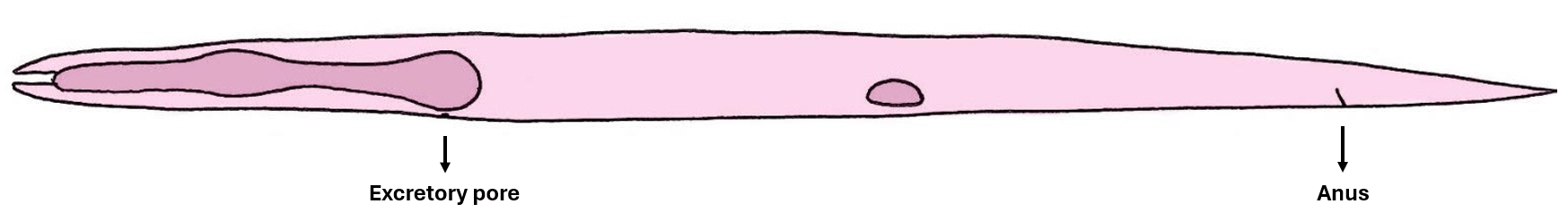
- In the head (anterior to the excretory pore) nuclei are divided in either ventrally or dorsally located;
- In the body (between excretory pore and anus) nuclei are located in the ventral side, in the lateral left side or in the lateral right side;
- In the tail (posterior to the anus) nuclei are located in the ventral side.
As an example: LL9 is a hyp7 nucleus that fuses to the syncytium during the L2 stage and is located in the lateral left side of the region of the body between the excretory pore and the anus; the cell that donates this nucleus to hyp7 is V1L.paa (anterior daughter of the anterior daughter of the posterior daughter of V1L), belonging to the seam cell lineage.