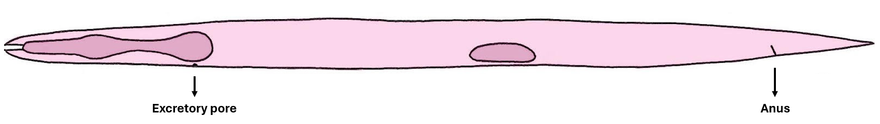
2D schematics of the nuclei contained in hyp7 by the end of the L2 stage
To allow for a clearer visualization, the nuclei are represented as split into three planes: lateral left (LL), medial and lateral right (LR).
The pharynx (in the head) and the gonad (in the mid-body) are represented as anatomical landmarks.
The nuclei are coloured according to the same colour-code as in the hyp7 nuclei lists. Hovering over a nucleus allows to visualize both the nucleus name and the cell name in an interactive fashion. A search bar also allows to highlight specific nuclei in the schematics.
Partial table of the nuclei contained in hyp7 by the end of the L2 stage
The organization of the table is the same as in the complete table of the hyp7 nuclei, except for the fact that each section of the body shows both the new nuclei acquired by the syncytium during the given stage and the total nuclei present by the end of the stage.
Loading workbook…
Download source XLSX file